CLID (Corneal Lesions Image Dataset)
The CLID dataset currently contains 30 images of the cornea’s surface, acquired during medical procedures with different lighting conditions and equipment, and including different types and levels of corneal lesions, thus increasing the variability of the data. The images were captured using a Topcon Sle corneal topographer, with 10 × magnification, in diffused lighting. The photos were obtained using an iPhone 5s smartphone connected to the Topcon Sle corneal topographer. After image capture, an experienced ophthalmologist, with a Ph.D. in applied sciences to surgery and ophthalmology, marked the area corresponding to the lesion in 140 the anterior eye segment in red, and the total area under analysis in blue, using a touch stylus pen. The specialist chose the red and blue colors of the marking because of the contrast with the background.
1. How To Use
Each image folder has two image files. One file representing the original image named "N_orig" (where N is the number of the image), and another file named N (where N is the number of the image). The files with the "_orig" suffix represent the original image, while the other file is the image marked by the ophthalmologist.
/Dataset
--/1
--/--/1
--/--/1_orig
--/2
--/--/2
--/--/2_orig
.
.
.
--/--/N_orig
1.1 Images
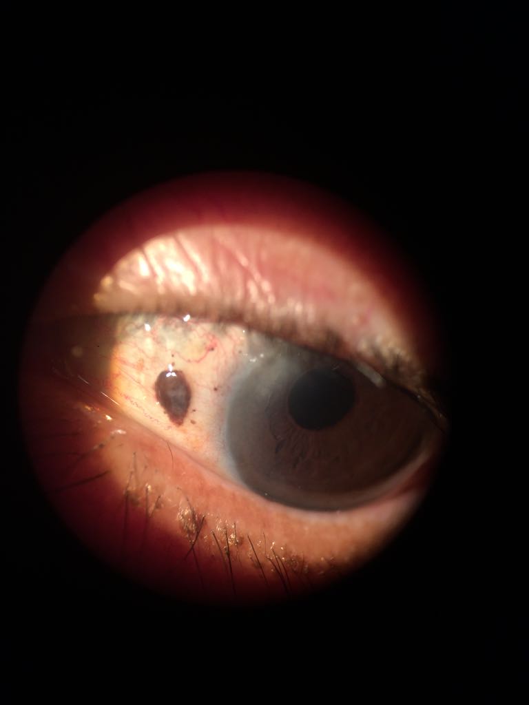
Original Image
Describe original image
Last updated 3 mins ago
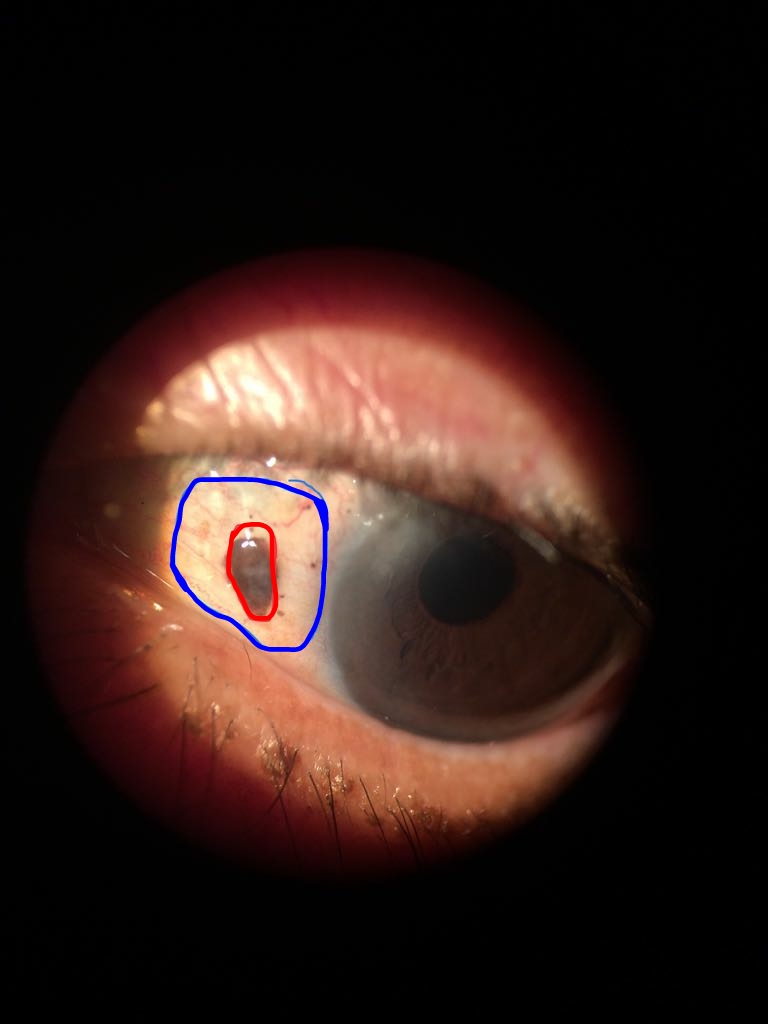
Marked Image
Describe marked image
Last updated 3 mins ago